
Inventor recalls eye imaging breakthrough
“It uses infrared light that’s barely visible compared to the bright flash of fundus photography [another common method of eye imaging] and provides a lot more information—three-dimensional rather than two-dimensional information—at higher resolution,” Huang says. The discovery earned him and his co-inventors slots in the National Inventors Hall of Fame in 2025 as well as the Lasker Award and the National Medals of Technology and Innovation in 2023.
Huang didn’t expect to change the paradigm of eye imaging when he began studying electrical engineering as an undergraduate at MIT, but he was interested in using an engineering mindset to contribute to medical advancements. That, he thought, could be his way to follow in the footsteps of his father, who was a family practitioner.
OCT emerged from his work as an MD-PhD student in the Harvard-MIT Program in Health Sciences and Technology. While studying ultrafast lasers at MIT under James Fujimoto ’79, SM ’81, PhD ’84, the Elihu Thomson Professor of Electrical Engineering, Huang was tasked with using the lasers to improve various ophthalmological tasks, including measuring the thickness of the cornea and retina.
Huang thought an approach known as interferometry, which could measure the time of flight down to one quadrillionth of a second, could improve thickness measurements to micrometer resolution. Huang’s experiments revealed that the technique was able to detect very faint signals arising from fine internal structures within the retina. Fujimoto and Huang realized the potential for inventing a new type of imaging and enlisted the help of Eric Swanson, SM ’84, who was using interferometry for intersatellite communications at Lincoln Laboratory, to develop an OCT machine for biological applications. Huang tested the new machine on several types of tissues accessed through Harvard Medical School and found it particularly successful in imaging retinal and coronary artery samples. He and his colleagues published their initial findings in Science in 1991, establishing OCT as a new imaging modality.
“Because of our ability to form collaborations with medical doctors and the more advanced technologies that were easily accessible at Lincoln Lab and MIT, we were able to make this new imaging technology take off when other people who were exploring around the same area were not able to demonstrate imaging results,” he says.
After the groundbreaking invention, Huang finished his academic and medical training as an ophthalmologist while Fujimoto and Swanson formed a startup company to ensure that the device got into medical offices.
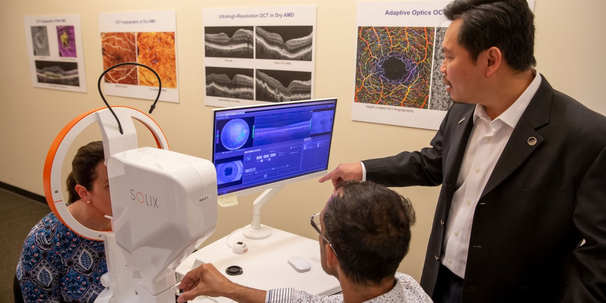
Over the next decades, Huang has continued to refine OCT for various applications. Today, as the director of research at Oregon Health and Science University’s Casey Eye Institute, he leads research groups exploring new ways to use OCT in techniques such as OCT angiography (imaging blood flow down to the capillary level) and OCT optoretinography (mapping the light response in retinal photoreceptor cells).
In addition to conducting research, he also sees patients and is the cofounder of GoCheck Kids, a digital platform for pediatric eye screening.
Huang credits his knack for innovation to his position at the nexus of diverse fields. “It’s hard for a pure medical doctor or a pure laser engineer to realize that there is an opportunity to invent a new device that solves a real problem in the clinic,” he says. “But it’s really easy when you have knowledge on both sides.”
24World Media does not take any responsibility of the information you see on this page. The content this page contains is from independent third-party content provider. If you have any concerns regarding the content, please free to write us here: contact@24worldmedia.com